What Is a Sequence Detection System? DNA, PCR, and How They Work Together
A sequence detection system is a lab instrument that finds and measures a chosen DNA or RNA target while PCR is happening. In simple words, PCR makes many copies of a tiny genetic target, and the sequence detection system watches that copying in real time through fluorescent light. That is why these systems are often called real-time PCR or qPCR systems.
DNA holds the genetic code, PCR copies a selected part of that code, and a sequence detection system reads the signal that proves the target is present. This is the quiet work behind many medical tests, research assays, food safety checks, forensic samples, and gene expression studies. When a result says a virus, mutation, gene, or bacterial marker was “detected,” a system like this may have helped create that answer. DNA is made from the four bases A, C, G, and T, and their order carries biological instructions.
What is a sequence detection system?
A sequence detection system is an instrument that detects a specific nucleic acid sequence usually DNA or RNA, by measuring fluorescent signals produced during PCR. It does not read the whole genome. It looks for a planned target sequence chosen by the assay designer.
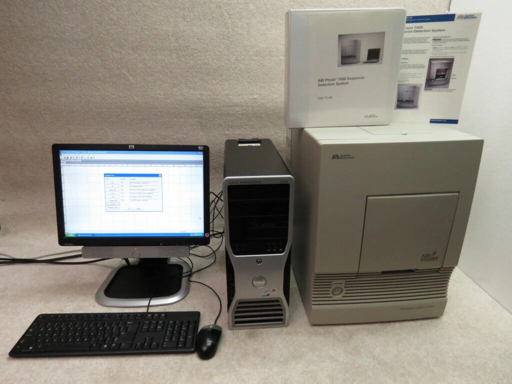
In many labs, the phrase “sequence detection system” refers to older Applied Biosystems real-time PCR instruments, such as the ABI PRISM or Applied Biosystems 7500 platforms. These systems combine thermal cycling, optical detection, and software analysis in one workflow. Systems such as the Applied Biosystems 7300, 7500, and 7500 Fast use fluorescent PCR chemistry to detect nucleic acid targets during real-time analysis. They can also support qualitative detection through end-point analysis or dissociation-curve analysis, depending on the assay setup.
The system has three main jobs. It heats and cools the sample so PCR can copy the target. It shines light into the reaction to measure fluorescence. Then it turns those light signals into curves, cycle threshold values, and positive or negative calls.
That may sound cold and mechanical, but the idea is beautifully simple. If the target DNA is there, the signal rises. If it is missing, the signal stays low or behaves like background noise.
How does DNA fit into sequence detection?
DNA is the target language that a sequence detection system is built to find. The system does not “see” traits, diseases, or organisms directly. It detects the DNA or RNA sequence linked to them.
DNA has a double-helix shape, with two linked strands wrapped around each other like a twisted ladder. Each strand has a sugar-phosphate backbone and bases called adenine, cytosine, guanine, and thymine. Those bases pair in a steady pattern. Adenine pairs with thymine, and cytosine pairs with guanine. The order of these bases is what scientists mean when they talk about a “sequence.” NHGRI explains that the ACGT code carries instructions in genes, and base pairing helps DNA copy itself with accuracy.
A sequence detection assay is built around that code. Scientists design short DNA pieces called primers to match the edges of the target. In probe-based assays, they also design a fluorescent probe that binds inside the target region. This is why the word “sequence” matters. The system is not just detecting any DNA. It is detecting a selected order of bases.
What is PCR?
PCR, or polymerase chain reaction, is a lab method that copies a selected DNA segment many times. It turns a tiny amount of genetic material into enough material for detection. PCR is widely used in research and clinical testing because it can amplify a selected DNA segment from a very small starting amount. The method depends on DNA polymerase, an enzyme that builds a new DNA strand from a template. PCR also needs primers because DNA polymerase can only add new bases from an existing starting point.
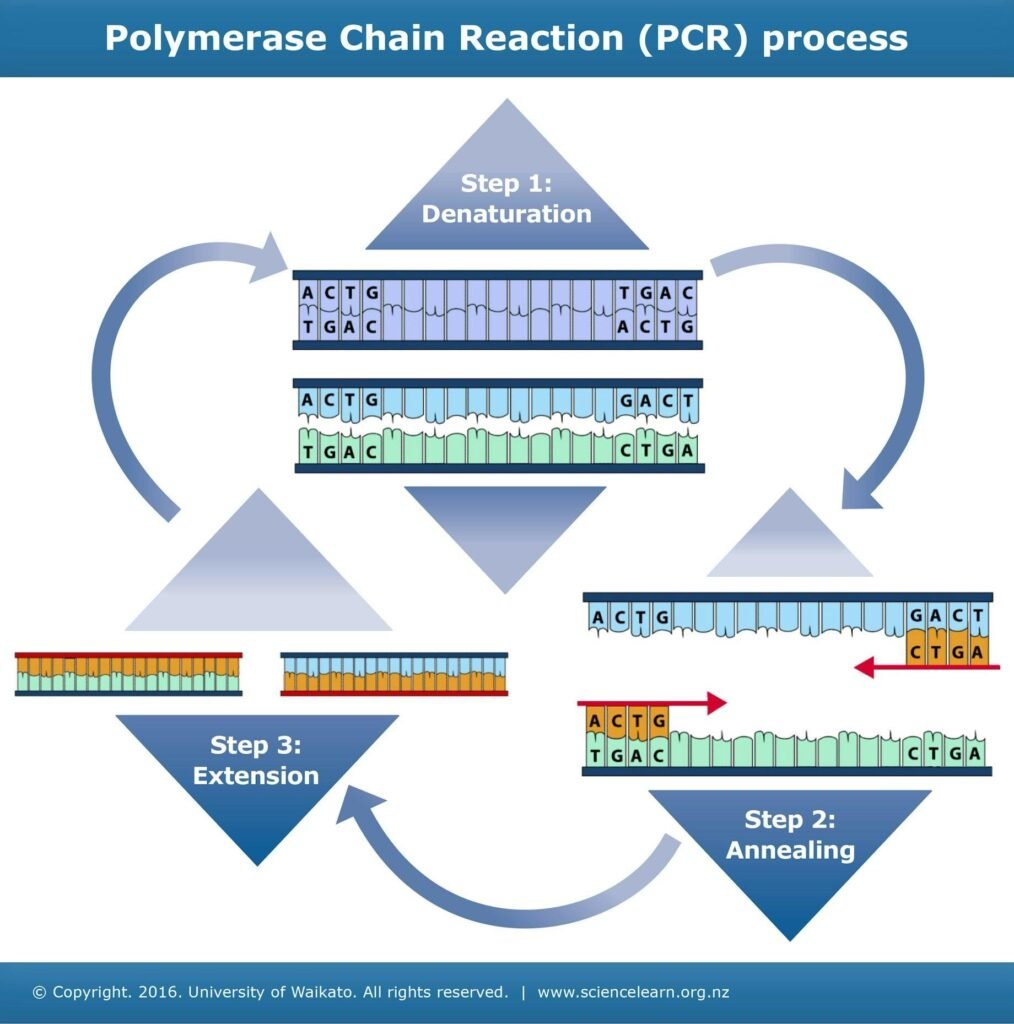
PCR works through repeated temperature cycles. Each cycle usually has three steps.
First, the DNA strands separate at a high temperature. Next, primers bind to their matching target sites as the sample cools. Then the enzyme extends the primers and builds new DNA. After one cycle, there are more copies than before. After many cycles, the target can grow from barely detectable to measurable.
This copying process is the reason PCR became one of the most used tools in molecular biology. It can find a specific genetic target even when the starting sample contains a messy mix of human DNA, microbial DNA, proteins, salts, and other material.
What is real-time PCR?
Real-time PCR measures DNA amplification while it is happening, instead of waiting until the end of the reaction. The instrument records fluorescence after each cycle and uses that rising signal to measure the target. Real-time PCR differs from conventional PCR because it detects amplified products while the reaction is still running, rather than checking the product only after amplification is finished. This reduces many post-PCR handling steps and uses fluorescent dyes or probes to track product formation cycle by cycle.
This is where sequence detection systems earn their name. A normal thermal cycler copies DNA, but a real-time sequence detection system copies and measures at the same time. The output is usually shown as an amplification curve. At first, the line stays flat because the fluorescence is too low to separate from background. Then, if the target is present, the signal rises. The cycle where that signal crosses a set threshold is often called Ct or Cq.
A lower Ct usually means more starting target was present. A higher Ct usually means less starting target was present. If no proper curve appears, the target may be absent, too low to detect, or affected by assay failure.
How do DNA, PCR, and a sequence detection system work together?
DNA provides the target, PCR copies the target, and the sequence detection system measures the copying through light. The three parts work as one chain: target, amplification, detection.
A sample might come from blood, saliva, tissue, food, water, or a swab. If the test is looking for DNA, the extracted DNA can go into PCR. If the target is RNA, as with many RNA viruses or gene expression tests, the RNA is usually converted into complementary DNA first. That method is called reverse transcription PCR, or RT-PCR.
Once the sample is prepared, primers and probes are added. The instrument then cycles the temperature again and again. As PCR creates new copies, fluorescent chemistry creates a signal. The system’s detector measures that signal after each cycle. Software plots the fluorescence and calculates whether the target crossed the threshold.
This workflow is powerful because it links biology to a readable number. A tiny genetic clue becomes a curve on a screen.
What happens inside the instrument during a run?
Inside the instrument, the reaction moves through heating, cooling, copying, light detection, and software analysis. Every part has to work cleanly for the final result to mean anything. The thermal block controls temperature. During denaturation, heat separates DNA strands. During annealing, primers attach to their matching sequence.
During extension, DNA polymerase builds new DNA. The optical system then measures fluorescence. Depending on the assay, that fluorescence may come from a dye that binds double-stranded DNA or from a probe that lights up only when a target sequence is copied.
The software turns fluorescence into an amplification plot. In a good positive reaction, the curve starts flat, rises through a clear growth phase, and may later level off when reaction components run low. Controls help the lab trust the run. A positive control should amplify. A no-template control should stay negative. An internal control can show whether extraction, chemistry, or sample inhibition affected the reaction. Without controls, a curve is just a curve. With controls, it becomes evidence the lab can read with more confidence.
What are the main detection chemistries?
Most sequence detection systems use either DNA-binding dyes or sequence-specific fluorescent probes. Both can detect PCR products, but they do it in different ways.
SYBR Green is a common dye-based method. It binds to double-stranded DNA and gives a much stronger fluorescent signal when attached to DNA than when free in solution. As more double-stranded PCR product forms, the signal rises.
That makes SYBR Green simple and often less costly. But because it binds to any double-stranded DNA, it can also detect primer dimers or off-target products. Labs often add melt-curve analysis after the run to check whether the product behaves like the expected target. TaqMan-style probe chemistry is more specific because it uses a fluorescent probe designed for the target sequence. SYBR Green, in comparison, detects accumulating PCR product through a double-stranded DNA dye.
A TaqMan probe carries a reporter dye and a quencher. When the probe is intact, the quencher keeps the reporter quiet. During PCR, the polymerase breaks down the probe as it copies the target. That separation lets the reporter dye shine.
This is why probe-based assays are often preferred when specificity matters, such as diagnostic testing, mutation detection, or multiplex assays where several targets are measured in the same tube.
What does Ct or Cq mean?
Ct or Cq is the PCR cycle number where fluorescence rises above a set threshold. It gives a rough picture of how much target was present at the start of the reaction.
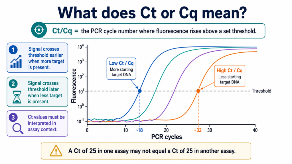
If a sample has a lot of target DNA, fluorescence rises earlier, so the Ct is lower. If a sample has very little target, it takes more cycles for the signal to rise, so the Ct is higher.
In qPCR, Ct or Cq refers to the number of cycles needed for fluorescence to cross a set threshold during the measurable growth phase of the reaction. Ct values should never be read alone without context. The assay, instrument, sample type, extraction method, controls, threshold setting, and lab rules all matter.
A Ct of 25 in one assay may not mean the same thing as a Ct of 25 in another. That is why trained staff follow set protocols instead of treating Ct as a universal number.
What is the difference between PCR, qPCR, and RT-PCR?
PCR copies DNA. qPCR copies and measures DNA in real time. RT-PCR starts with RNA, turns it into complementary DNA, and then amplifies it.
These terms often get mixed up, especially because “RT-PCR” can mean reverse transcription PCR, while “real-time PCR” is also sometimes shortened in casual speech. That confusion became common during public discussions of viral testing.
A clean way to remember it is this:
PCR asks, “Can we copy this target?”
qPCR asks, “How much target appears as copying happens?”
RT-PCR asks, “Can we detect an RNA target after converting it to DNA?”
Real-time RT-PCR combines both ideas. It starts with RNA, converts it to DNA, then measures amplification in real time. This method is widely used for RNA viruses and gene expression work. Real-time PCR is used across many biomedical applications, including the detection of emerging viruses, because it can identify selected genetic targets quickly and with high sensitivity.
What is a sequence detection system used for?
Sequence detection systems are used to detect, measure, and compare specific DNA or RNA targets. Their uses span health care, research, agriculture, food safety, environmental testing, and forensic science.
In medical labs, these systems can help detect pathogens, measure viral load, study cancer markers, or test genetic variants.
In research labs, they often measure gene expression, copy number, or small changes in DNA. In food and water testing, sequence detection can identify microbial contamination. In forensic work, PCR-based methods can help analyze tiny DNA samples, though the workflows differ from clinical qPCR.
The power comes from specificity. A lab can design an assay for one short target sequence and ask whether it exists in a sample.
That makes sequence detection useful when the answer must be more exact than “something grew in culture” or “something looks suspicious under a microscope.”
Why are sequence detection systems so sensitive?
They are sensitive because PCR copies the target many times, and the instrument measures signal during each cycle. A small starting amount can become detectable after repeated amplification.
PCR is often described as exponential in its early phase. In an ideal reaction, each cycle roughly doubles the amount of target. Real reactions do not stay perfect forever, but early amplification can still turn a tiny starting signal into a clear curve. That sensitivity is both a strength and a weakness. It helps labs detect low-level targets. It also means contamination can cause false positives if workflows are sloppy.
A few stray DNA molecules from a previous amplification run may be enough to create a signal. Good labs separate sample preparation, reaction setup, and post-amplification areas. They also use filtered tips, negative controls, and strict cleaning habits.
The system can detect small signals, but human technique protects the meaning of those signals.
How accurate is a sequence detection system?
A sequence detection system can be highly accurate when the assay is well designed, the sample is clean, controls pass, and staff follow validated protocols. The instrument is only one part of the result. Accuracy begins before the run. The sample must be collected, stored, and extracted properly. Bad sampling can cause a false negative even if the instrument works perfectly.
The primers and probes also matter. They must match the target and avoid close matches to unrelated sequences. Assay design becomes even more delicate when the target organism mutates or when the sample contains many related species. For research settings, the MIQE guidelines are often used to support better reliability and transparency in qPCR studies. They set out the minimum information needed to judge qPCR experiments and include a reporting checklist for assay details, sample handling, controls, and analysis.
That tells us something practical: qPCR is not magic. It is a measured process that needs careful reporting, controls, and repeatable methods.
What can go wrong during sequence detection?
False positives, false negatives, weak curves, primer dimers, poor extraction, inhibitors, and software threshold errors can all affect results. Most problems come from sample quality, assay design, contamination, or interpretation. A false positive can happen if target DNA contaminates a reaction. Because PCR is so sensitive, even small contamination can matter. A false negative can happen when the sample has too little target, when extraction fails, when inhibitors block the enzyme, or when the target sequence has changed enough that primers or probes bind poorly.
Weak curves may appear when the target is near the limit of detection. Strange curves may point to bubbles, evaporation, optical problems, or nonspecific amplification.
SYBR Green assays need extra care because the dye detects any double-stranded DNA. Probe assays add specificity, but they still depend on good design and clean technique. This is why labs rarely trust a single number without controls. A reliable answer comes from the whole pattern.
How is a sequence detection system different from DNA sequencing?
A sequence detection system checks for a known target sequence. DNA sequencing reads the order of bases in a DNA molecule. This is a key difference. A sequence detection system is like asking, “Is this sentence in the book?” Sequencing is like reading the pages. qPCR is usually faster, cheaper per target, and easier for routine testing when the lab knows what it is looking for.
Sequencing gives broader information and can find new changes, but it often needs more analysis, more time, and different equipment.
For example, qPCR can detect whether a pathogen marker is present. Sequencing can show the exact genetic code of that pathogen region, which may help track variants or study mutations. Neither method replaces the other. They answer different questions.
Why do labs still use sequence detection systems when sequencing exists?
Labs still use sequence detection systems because they are fast, targeted, sensitive, and practical for routine testing. When the target is known, real-time PCR can give clear answers without reading extra genetic material. A hospital lab may need to know whether a specific pathogen is present. A research lab may need to compare expression of one gene across 100 samples. A food lab may need to screen for one contamination marker. In those cases, sequencing may be more information than the lab needs.
Real-time PCR systems can also process many samples in plates, often 96 wells or more, depending on the instrument. Older Applied Biosystems 7500 systems were built as integrated platforms for detecting and measuring nucleic acid sequences. Speed matters too. Some real-time PCR systems support fast cycling. The 7500 Fast system, for example, was designed for high-speed thermal cycling and shorter run times compared with standard cycling workflows.
What does the software show after the run?
The software usually shows amplification curves, Ct or Cq values, baseline settings, thresholds, melt curves for dye assays, and result calls. These outputs help the lab decide whether each sample is positive, negative, invalid, or needs repeat testing. The amplification plot is the main view. It shows fluorescence against PCR cycle number. A clean positive sample has a curve that rises in the expected shape. A negative sample should stay flat, similar to the no-template control.
For SYBR Green assays, melt-curve analysis can help check whether the reaction produced one main product. A single expected melt peak supports specificity. Extra peaks may suggest nonspecific products or primer dimers. The software helps, but it does not replace judgment. Someone still has to review controls, curve shape, threshold placement, and assay rules.
What should beginners remember about sequence detection?
Beginners should remember that sequence detection is not the same as reading a whole genome. It is a targeted test that asks whether a chosen DNA or RNA sequence is present and, in many assays, how much is there. The workflow has a simple story:
- DNA or RNA is extracted from a sample.
- PCR copies the target region.
- Fluorescent chemistry creates a signal.
- The instrument measures that signal.
- Software turns it into curves and values.
- The lab reads the result with controls.
Once that story clicks, the whole system feels less mysterious. The instrument is not “finding disease” by itself. It is detecting a molecular signal that a trained assay connects to a biological question.
What is the future of sequence detection systems?
Sequence detection systems are likely to stay central in labs because they answer focused genetic questions quickly. Newer instruments may become faster, smaller, more automated, and easier to connect with lab software, but the core idea remains the same. DNA carries the code. PCR copies the target. Fluorescence reports the copying. The system reads the signal.
That simple chain is why real-time PCR has lasted so long in science and medicine. Even as sequencing grows, targeted detection still has a place wherever speed, sensitivity, and clear yes-or-no answers matter.
A good sequence detection result is more than a line on a screen. It is a carefully built answer from chemistry, biology, optics, software, and human care.