How Do Labs Validate a Sequence Detection Assay?
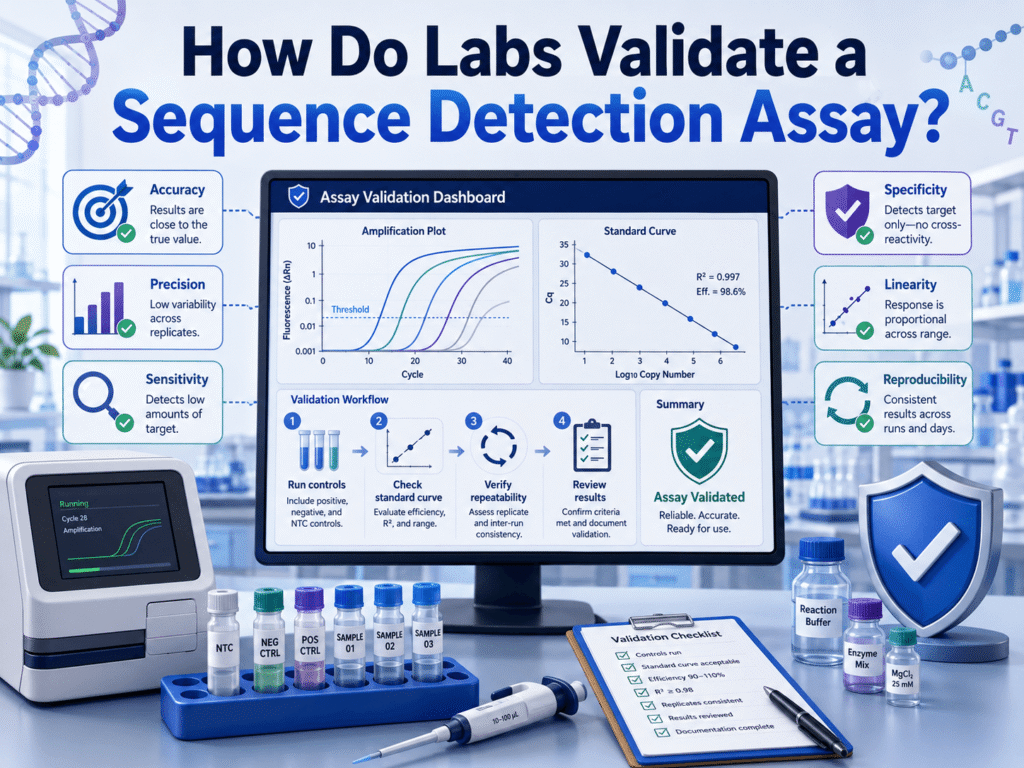
A lab validates a sequence detection assay by proving, with planned experiments, that the test can find the right DNA or RNA target in the right sample type, at the claimed detection limit, without giving too many false positives or false negatives.
Validation checks accuracy, precision, analytical sensitivity, analytical specificity, reportable range, controls, contamination risk, and clinical performance before patient or research results are trusted. Under CLIA-style expectations, analytical validation covers accuracy, precision, analytical sensitivity, analytical specificity, reportable range, reference interval, and any other performance feature needed for that laboratory’s test system.
That may sound technical, but the concern behind it is very human. A sequence assay result can guide treatment, infection control, cancer care, inherited disease workups, food safety actions, or research decisions. One weak validation step can turn a clean-looking report into a misleading answer. Good labs do not ask, “Did the assay work once?” They ask, “Can it keep giving the right answer when real samples, low target levels, inhibitors, operators, instruments, and near-neighbor sequences challenge it?”
What is a sequence detection assay?
A sequence detection assay is a laboratory test designed to detect a known DNA or RNA sequence in a sample, and its validation depends on understanding how sequence detection works from target amplification to signal interpretation. The target may come from a virus, bacterium, fungus, parasite, human gene, tumor mutation, transgene, or environmental organism.
These assays can use PCR, RT-PCR, qPCR, digital PCR, Sanger sequencing, targeted NGS, hybridization, or other molecular methods. Molecular testing guidance usually covers both amplified and non-amplified nucleic acid methods, along with sequence selection, inhibitors, false-positive control, reporting, and quality assurance.
A simple example is a PCR assay that detects a viral gene. A more complex example is a multiplex panel that checks many microbial targets in one reaction. For multiplex molecular tests, ISO 21474-2:2022 gives general requirements for validation and verification of tests that identify two or more nucleic acid targets at the same time.
What is assay validation in a lab?
Assay validation is the documented process of showing that a test performs well enough for its intended use. It answers one central question: can this assay produce dependable results for the sample types, targets, instruments, staff, and workflow used in this lab?
Validation is broader than a one-time setup check. It usually includes a written plan, selected reference materials, acceptance criteria, wet-lab testing, data review, approval, staff training, and post-launch monitoring.
Validation and verification give laboratories a structured way to plan studies, run experiments, review data, document findings, and keep records for new testing methods.
In molecular assays, the main goal is: the lab must show that the assay detects what it claims to detect, under the conditions in which the lab will use it.
Validation vs verification: what is the difference?
Validation is used when the lab develops a test, modifies a cleared test, changes the intended use, or creates a new workflow. Verification is used when the lab adopts an already cleared or approved method and confirms that it performs as expected in that local setting.
Before a new test enters clinical use, labs usually verify FDA-cleared or FDA-approved methods, while laboratory-developed methods need full validation.
This difference is crucial because a manufacturer’s package insert does not automatically prove the assay works in every lab. A local lab may use different staff, extraction instruments, thermocyclers, specimen transport conditions, software versions, or sample mixes.
For example, if a lab uses an FDA-cleared respiratory PCR kit exactly as written, it may verify accuracy, precision, and reportable conditions. If the same lab changes the specimen type, adds pooling, changes extraction chemistry, or modifies the cutoff, it may need a deeper validation.
What is the first step in validating a sequence detection assay?
The first step is to define the assay’s intended use in writing. The lab should state what target is detected, which specimen types are allowed, whether the result is qualitative or quantitative, who the test is for, and how the result will be reported.
This step shapes every experiment that follows. A test for “SARS-CoV-2 RNA in nasopharyngeal swabs” is not automatically valid for saliva, wastewater, blood, or formalin-fixed tissue. A tumor mutation assay valid for a hotspot region is not valid for the whole gene unless the validation supports that claim.
For multiplex assays, CLSI MM17 covers method selection, specimen and reagent assessment, analytical validation of laboratory-developed and modified IVD assays, amplification technologies, data analysis, and reporting.
A good intended-use statement usually names the target, technology, specimen, extraction method, instrument, result type, reportable units, cutoff, and user setting.
How do labs choose samples for validation?
Labs choose samples that represent the real world as closely as possible. That means positive samples, negative samples, weak positives near the detection limit, different specimen types, interfering substances, extraction controls, and organisms or sequences that could be confused with the target.
Clinical samples are best when enough are available. Contrived samples may be used when positives are rare, but the lab should prepare them in the right sample matrix. For infectious disease assays, this may mean spiking quantified viral or bacterial material into swab medium, sputum, stool, blood, plasma, or another validated matrix.
FDA-style molecular diagnostic templates for respiratory tests have commonly focused on limit of detection, clinical evaluation, inclusivity, and cross-reactivity as key validation data categories.
The closer the validation material is to patient material, the more useful the study becomes. Purified DNA in water may show that primers work, but it does not fully challenge extraction, inhibitors, transport medium, sample viscosity, or matrix effects.
How do labs test analytical sensitivity?
Analytical sensitivity asks how little target the assay can detect. In sequence detection assays, this is usually measured as the limit of detection, often shortened to LoD.
A lab tests LoD by preparing a dilution series of known target material and running multiple replicates at each level. The claimed LoD is often the lowest concentration detected with a defined hit rate, such as 95%, depending on the method and regulatory setting.
According to FDA’s molecular diagnostic EUA, LoD testing use all parts of the test system, from sample preparation and extraction through detection.
Detection is not only about primers and probes. Extraction losses, elution volume, inhibitors, transport media, and instrument thresholds can all change sensitivity.
For qPCR, a strong validation usually checks PCR efficiency, linear dynamic range, LoD, and precision.
How do labs confirm analytical specificity?
Analytical specificity checks whether the assay detects only the intended target and avoids false positives from similar organisms, genes, or background material.
Specificity testing in infectious disease assays often has two sides: inclusivity and exclusivity. Inclusivity asks whether the assay detects known variants, strains, genotypes, or subtypes of the target. Exclusivity asks whether it avoids cross-reactivity with near neighbors and common organisms found in the same specimen type.
For SARS-CoV-2-style molecular templates, inclusivity and cross-reactivity are usually treated as core specificity checks.
In silico analysis is often used first. The lab compares primer and probe sequences against databases to see whether mismatches could block detection or whether unrelated organisms share enough sequence similarity to create a false signal. Wet testing may then be done using cultured organisms, extracted nucleic acid, synthetic material, or clinical samples.
For genetic assays, specificity may include checking pseudogenes, homologous regions, repetitive DNA, nearby variants under primers, and off-target amplicons.
How do labs measure accuracy?
Accuracy checks whether the new assay agrees with the true or accepted result. Labs usually compare results against a reference method, a previously validated assay, sequencing, reference materials, proficiency samples, or well-characterized clinical samples.
For qualitative sequence detection, accuracy often becomes positive agreement and negative agreement. If 100 known positive samples are tested and 98 are detected, the positive agreement is 98%. If 100 known negative samples are tested and 99 are negative, the negative agreement is 99%.
Lab-developed molecular assays usually require core checks such as accuracy, precision, reportable range, reference interval, analytical sensitivity, and analytical specificity.
Accuracy should include weak positives when possible. A validation set full of strong positives can make an assay look better than it really is. Real failures often happen near the cutoff, in low-copy samples, degraded samples, or samples with inhibitors.
How do labs test precision and reproducibility?
Precision checks whether the assay gives the same result when repeated. Reproducibility checks whether the result holds across days, operators, reagent lots, instruments, and runs.
A lab may test the same positive, low-positive, and negative material over several days. It may include different staff members and instruments if those will be used after launch. For qPCR, precision can be measured through Cq or Ct variation. For qualitative assays, the lab may track percent agreement across replicates.
Precision is one of the main performance characteristics labs need to establish when they modify an FDA-cleared test or create a laboratory-developed test along with accuracy, analytical sensitivity, interferences, analytical specificity, and reportable range where relevant.
Low positives are especially useful here. A strong positive may repeat beautifully, while a sample near the LoD may expose random failures.
How do labs set the cutoff or threshold?
Labs set the cutoff by reviewing validation data and deciding where positive, negative, and sometimes indeterminate results should fall. In qPCR, this may involve Ct or Cq values, amplification curve shape, replicate rules, internal control behavior, and background noise.
The cutoff should separate true signal from noise without hiding weak true positives. That balance is not always easy. A cutoff that is too loose may raise false positives. A cutoff that is too strict may miss low-level target.
For qPCR and RT-qPCR methods, method development guidance commonly links sensitivity to LoD and lower limit of quantification for quantitative use, while also using no-template controls to check nonspecific amplification and contamination.
For sequencing assays, the cutoff may include read depth, allele fraction, base quality, mapping quality, strand balance, contamination metrics, and variant-calling filters. These thresholds should be tested, not guessed.
What controls are used during validation?
Controls show whether each part of the assay worked and whether contamination occurred. A sequence detection assay usually needs positive controls, negative controls, no-template controls, extraction controls, internal controls, and sometimes inhibition controls.
A no-template control contains the assay reagents but no target template. It checks contamination or nonspecific amplification. No-template negative controls are considered as nuclease-free molecular-grade water or buffer used to monitor nonspecific amplification, cross-contamination during setup, and nucleic acid contamination of reagents.
An extraction control travels through extraction and amplification. It can reveal contamination or extraction failure that a late-added control may miss. An internal control is often added to each sample to show that extraction and amplification were not blocked.
NGS assays rely on no-template controls to detect background contamination from reagents, plastics, water, barcode bleed-through, or library preparation steps.
How do labs test interference and inhibition?
Labs test interference by adding substances that may appear in real samples and checking whether they block detection or create false signals.
For respiratory samples, this may include mucus, blood, nasal sprays, throat medications, or transport media. For stool, inhibitors may include bile salts and complex organic material. For blood or plasma, hemoglobin, anticoagulants, lipids, and high human DNA background may matter.
Inhibitors and interfering substances are a standard part of molecular infectious disease assay validation.
Inhibition is a common problem in nucleic acid assays. A sample can contain the target, but the reaction may fail because enzymes are blocked or extraction is poor. Internal controls help catch this, but validation should also show how the assay behaves when inhibitors are present.
How is reportable range validated?
Reportable range is the span of results the lab can report with confidence. For qualitative assays, it may be limited to detected, not detected, invalid, or indeterminate. For quantitative assays, it includes the range over which the reported number is accurate and precise enough.
For qPCR, this often requires a dilution series across several concentrations. The lab checks linearity, efficiency, precision, and lower and upper limits. PCR efficiency, linear dynamic range, LoD, and precision are assay performance characteristics for qPCR.
Sanger or targeted sequencing – reportable range may refer to the exact exons, codons, genes, hotspots, or genomic positions covered by the assay. In Sanger sequencing and pyrosequencing, reportable range can be the nucleotide mutations identified in the codons or exons of interest.
A lab should not report beyond the validated region. If an assay covers exon 12, the report should not imply the full gene was assessed.
How do labs validate multiplex sequence detection assays?
Multiplex validation is harder because many targets are tested at once. The lab must show that one target does not interfere with another, that mixed infections or mixed templates can be detected, and that strong positives do not hide weak positives.
Multiplex nucleic acid assay validation requires attention to sample preparation, reference materials, quality control materials, data analysis, and reporting.
Multiplex assays also need inclusivity and exclusivity for each target. A respiratory panel with 20 targets is not one validation question; it is many linked questions. The lab must think about cross-reactivity, competition for reagents, probe overlap, spectral bleed-through, primer dimers, and interpretation rules.
For syndromic panels, clinical interpretation also needs care. Detecting a sequence does not always prove active disease. The report language should match what the assay can support.
How do labs validate NGS-based sequence detection?
NGS validation includes all the usual molecular questions, plus sequencing-specific checks. The lab reviews library preparation, target enrichment, read depth, coverage uniformity, base quality, mapping, variant calling, contamination, index hopping, bioinformatics settings, and report filters.
For targeted NGS, no-template and negative controls can reveal reagent or workflow contamination. AMP/CAP NGS validation guidance recommends including a no-template control in targeted NGS library preparation to check reagent contamination.
NGS validation also needs well-characterized reference samples. For human variants, labs may use Genome in a Bottle-type materials, cell lines, synthetic controls, orthogonal sequencing, or known clinical specimens. For microbial sequencing, labs may use mock communities, quantified organisms, reference genomes, and contamination controls.
The bioinformatics pipeline is part of the assay. Changing software, databases, alignment settings, variant callers, or filtering rules can change results and may require revalidation or partial validation.
How do labs document assay validation?
Labs document the validation plan, raw data, calculations, deviations, acceptance criteria, summary, approval, and final standard operating procedure.
The validation file should tell a future reviewer what was tested, why it was tested, what materials were used, who performed the work, how results were analyzed, what passed, what failed, and what limits remain.
Validation and verification records should be easy to retrieve when inspectors or assessors ask to review them.
Good documentation also protects the lab. Months later, when a result is questioned, a clear validation file can show the assay’s claimed use, sample types, cutoff, LoD, known limitations, and control rules.
When does a lab need to revalidate a sequence detection assay?
A lab may need revalidation, or at least a partial validation, when something changes that could affect results. This can include a new extraction kit, new instrument, new reagent lot type, new specimen type, new primer or probe sequence, new cutoff, new software, new target region, or new reporting claim.
Re-checking inclusivity may also be needed in infectious disease assays; when the target organism changes. Viral mutations under primers or probes can reduce detection. Public sequence databases can help monitor this risk, but wet testing may still be needed when a major mismatch appears.
NGS pipeline updates are a common trigger for NGS. A minor user-interface change may not affect results, but a new aligner, reference genome, variant caller, database, or filter can change variant calls.
The safest rule is practical: if the change could alter the result, the lab should document why no study was needed or perform a study scaled to the risk.
What makes a validation study strong?
A strong validation study matches the assay’s real use. It includes the right samples, weak positives, true negatives, near-neighbor sequences, controls, multiple runs, and clear acceptance criteria.
It also admits the assay’s limits. No sequence detection assay is perfect. A negative result may mean no target was detected, not that the organism or variant is absent in every possible sense. Poor collection, low target level, inhibitors, degradation, or sequence changes can all affect results.
A strong report uses careful wording. It does not overstate the clinical meaning of a molecular signal. It tells users what the assay detects, what it does not detect, which specimens are accepted, and when repeat testing or another method may be needed.
A validated assay is a promise the lab must keep
Labs validate sequence detection assays to build trust before results leave the bench. The process starts with a clear intended use, then moves through sensitivity, specificity, accuracy, precision, interference, controls, reportable range, and documentation.
PCR, qPCR, dPCR, Sanger sequencing, and NGS, the details differ, but the core promise stays the same: the assay should detect the right sequence, in the right sample, with known limits.
A well-validated assay does more than pass inspection. It gives clinicians, researchers, and patients a result they can act on with more confidence. That is the real value of validation: fewer guesses, cleaner decisions, and a lab report that means exactly what it says.