Why Is Sample Preparation Important in Sequence Detection?
Sample preparation is important in sequence detection because even the best instrument can only read what the sample allows it to read. If the specimen is poorly collected, degraded, contaminated, diluted, or full of PCR inhibitors, the final result can become weak, unclear, false-negative, or misleading. Sample preparation protects the truth inside the sample.
A sequence detection system may look advanced on the outside. It may use qPCR, digital PCR, Sanger sequencing, next-generation sequencing, or isothermal amplification. Still, every result starts with the same quiet step: getting the right biological material into a clean, stable, test-ready form.
That step can decide whether a lab sees a real pathogen, mutation, variant, or genetic marker; or misses it completely. Good sample preparation does not just support the test. It shapes the quality of the answer.
What is sample preparation in sequence detection?
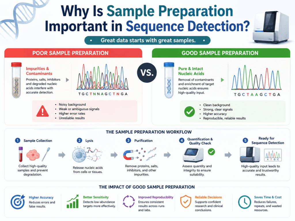
Sample preparation is the process of turning a raw specimen into material that a sequence detection system can test reliably. It may include collection, labeling, storage, transport, cell lysis, nucleic acid extraction, purification, concentration, and quality checks.
A raw sample is rarely ready for detection as it is.
Blood contains cells, proteins, enzymes, and compounds that may interfere with testing. Saliva can contain mucus, food traces, microbes, and human DNA. Tissue may need careful cutting, digestion, or preservation. A swab may carry only a small amount of target material.
The goal is to protect the DNA or RNA that needs to be detected while removing anything that can block the reaction.
In PCR-based sequence detection, the sample must contain enough target nucleic acid for amplification. In sequencing, the sample may need extra checks for purity, fragment size, and concentration before library preparation can begin.
A lab may have a powerful machine, well-designed primers, and trained staff. Poor sample preparation can still weaken the whole workflow before the instrument even starts.
Why does sample preparation affect sequence detection results?
Sample preparation affects sequence detection results because the test depends on the quality, purity, and amount of nucleic acid present in the tube. Bad preparation can reduce target recovery, damage RNA or DNA, introduce contamination, or leave behind substances that block amplification.
Sequence detection is a chemistry, the reaction needs target material, enzymes, primers and probes, buffers, and stable conditions. When the prepared sample is clean and well-preserved, the detection system has a fair chance to find the target.
When the sample is messy or degraded, the system may struggle.
A low-quality sample can produce a high Ct value in qPCR, uneven read depth in NGS, poor signal in Sanger sequencing, or failed amplification in an isothermal assay. These failures may look like instrument problems, but the root cause often sits earlier in the process.
Preanalytical factors in molecular testing can affect the outcome before amplification or sequencing even begins. Sample preparation is where the lab protects sensitivity, specificity, repeatability, and confidence.
What happens when a sample is collected poorly
Poor sample collection can leave the test with too little target material, the wrong material, or material that has already started to degrade. In infectious disease testing, this can lead to a false-negative result even when the person truly has the infection.
Collection is the first point where the test can go right or wrong.
A nasal swab that barely touches the correct site may not pick up enough viral RNA. A blood tube filled below the required volume may disturb the ratio between blood and additives. A tissue sample taken from a low-tumor area may miss the mutation that the clinician wants to find.
The issue is not always carelessness. Some targets are hard to capture. Some samples have low organism load. Some patients are tested late, after the amount of target has dropped.
Still, collection technique is important.
The specimen should match the test’s intended use. A respiratory virus assay may need a respiratory specimen. A mutation assay may need enough tumor content. A bloodstream infection test may need proper blood volume.
Even with sensitive RT-PCR methods, false-negative results can occur from poor specimen collection or poor handling. When the wrong specimen enters the workflow, even perfect extraction and perfect amplification may produce the wrong answer.
Why is nucleic acid extraction such a key step?
Nucleic acid extraction is key because it releases DNA or RNA from cells, viruses, bacteria, or tissue and separates it from proteins, membranes, salts, enzymes, and other reaction-blocking material. Without clean extraction, detection may fail even when the target is present.
Extraction sits near the heart of sample preparation.
The test cannot detect genetic material that remains trapped inside cells or viral particles. Lysis breaks open those structures. Purification then separates nucleic acid from unwanted sample components.
Different sample types need different handling.
Blood, stool, sputum, formalin-fixed tissue, plasma, and environmental samples behave differently. Stool often contains compounds that block PCR. Sputum can be thick and difficult to process. RNA from respiratory samples may degrade quickly if not protected.
A good extraction method gives the lab enough clean DNA or RNA for the next step.
Too little extraction can lower sensitivity. Harsh extraction can fragment nucleic acid. Incomplete purification can leave inhibitors behind. Cross-contamination during extraction can create false-positive results.
Collection, transport, preparation, storage, and nucleic acid isolation all sit inside molecular test quality, not outside it. The instrument may report the final signal, but extraction often decides how strong that signal can be.
How does sample preparation help prevent false-negative results?
Sample preparation helps prevent false-negative results by preserving target nucleic acid, removing inhibitors, and concentrating enough material for detection. A false-negative result can happen when the target exists in the patient or specimen but does not survive the workflow in testable form.
False negatives are dangerous because they feel final.
A patient may be told an infection was not detected. A tumor mutation may be missed. A food safety sample may appear clean. A genetic marker may go unseen.
Many false negatives begin with weak input.
The sample may contain too little target. RNA may break down during storage. Extraction may recover only a small fraction of the material. A compound in the specimen may block the enzyme that should copy the target sequence.
PCR tests check for small amounts of genetic material in samples such as blood, saliva, mucus, or tissue. That makes them powerful, but they still need usable material in the reaction.
Internal controls help catch some of these problems. A control can show whether extraction worked, whether amplification was blocked, or whether the run failed. Yet controls do not fix a poor sample. They only warn the lab that something may be wrong.
Strong sample preparation gives the test a better chance to detect low-level targets before they disappear into noise.
How does sample preparation reduce false-positive results?
Sample preparation reduces false-positive results by lowering the chance that outside DNA, RNA, amplified product, or sample mix-ups enter the test. False positives can happen when the system detects material that came from contamination rather than the true specimen.
Sequence detection methods can be extremely sensitive. That sensitivity is useful when the target is rare. It also creates risk. A tiny amount of contaminating nucleic acid can be copied millions of times during amplification.
Contamination can come from many places. It may come from another patient sample, a previous PCR product, a shared pipette, an opened tube, a reagent, or an unclean work area. In NGS workflows, barcode hopping, index mix-ups, or carryover can also confuse interpretation when controls are weak.
Good sample preparation uses separation and clean handling.
Pre-amplification and post-amplification areas should not mix. Reagents should be handled carefully. Negative controls should travel through the process. Tubes should be opened and closed with care. Staff should avoid moving amplified material back into clean spaces.
Routine monitoring can help laboratories find patterns that may point to false-positive molecular testing problems. A clean workflow protects trust in positive results.
Without that protection, a “detected” result may not come from the sample at all.
Why does RNA need extra care during sample preparation?
RNA needs extra care because it breaks down more easily than DNA. RNases are common in the environment, on skin, and in many biological samples, so RNA-based sequence detection needs fast handling, proper storage, and protective reagents.
RNA testing is common in viral detection, gene expression analysis, transcriptomics, and some cancer assays.
The problem is that RNA is fragile.
A sample left too long at room temperature may lose part of its target. Freeze-thaw cycles can damage RNA. Poor storage can make an originally strong sample look weak. In gene expression work, delayed handling can even change the biological pattern being measured.
This is one reason pre-test handling matters so much in RNA workflows.
The lab may need RNA-stabilizing media, cold-chain transport, RNase-free tubes, careful extraction, and fast processing. In RT-qPCR, RNA must also be converted into complementary DNA before amplification, which adds another step where quality can change.
NAATs can detect very small amounts of viral RNA, but only when that RNA reaches the test in a usable state. DNA is more stable, but RNA often tells a more time-sensitive story.
If that story is not protected early, the final result may be only a faint shadow of the original sample.
## How do inhibitors affect sequence detection?
Inhibitors affect sequence detection by blocking the enzymes used in amplification or library preparation. These substances can delay the signal, weaken amplification, reduce sequencing quality, or cause a complete test failure.
Many sample types carry natural inhibitors.
Blood may contain heme. Stool may contain bile salts and complex organic material. Soil and water samples may contain humic substances. Sputum can contain mucus and cellular debris. Some collection devices or preservatives can also interfere if they do not match the assay.
A sequence detection system may not always know the difference between “no target” and “target blocked by inhibitors.”
That is the scary part.
A qPCR curve may appear late or flat. An isothermal test may fail to amplify. An NGS library may perform poorly. If no internal control is used, the result may be wrongly treated as negative. This is why PCR inhibition remains a real problem across qPCR, digital PCR, and sequencing workflows.
Sample preparation reduces this risk by washing, purifying, diluting, or separating nucleic acid from inhibitory substances.
Dilution can sometimes reduce inhibition, but it may also dilute the target. Better extraction and purification usually give a safer path, especially when the target is already low.
A clean sample gives the enzyme room to work.
Why does sample storage and transport affect detection?
Storage and transport affect detection because heat, time, light, enzymes, and repeated freeze-thaw cycles can damage nucleic acids before testing begins. Even a well-collected sample can lose value if it is stored or shipped under poor conditions.
The sample does not pause while it travels.
Cells can break down. RNA can degrade. Bacteria can overgrow. Enzymes can remain active. A specimen that was usable at collection may become weak by the time it reaches the lab.
Transport media, temperature, and timing all shape final quality.
Some samples need refrigeration. Some need freezing. Some need stabilizing media. Some must be processed within a narrow time window. The right choice depends on the test, target, specimen type, and lab method.
This is especially relevant when samples come from remote clinics, home collection kits, field studies, or multi-site trials.
A central lab may run the same test with the same instrument every day. Yet if one clinic ships samples cold and another ships them warm, the data may not be truly comparable.
The final result often depends on basic pre-test details such as specimen collection, transport, preparation, and storage.
Good transport does not make a poor sample perfect, but poor transport can ruin a good one.
What role does sample quantity play in sequence detection?
Sample quantity plays a major role because detection systems need enough starting material to find the target with confidence. Too little input can raise Ct values, reduce sequencing depth, increase dropout, or make rare variants harder to detect.
Quantity is not only about volume.
A tube may contain enough liquid but too few cells. A swab may look wet but carry little target. A biopsy may contain mostly healthy tissue when the assay needs tumor DNA. Plasma may contain very low amounts of circulating tumor DNA.
Low input creates uncertainty.
In qPCR, low target levels can appear near the limit of detection. In NGS, low input may cause uneven coverage or missed variants. In digital PCR, too little target may limit the strength of the final count.
Too much input can also cause problems.
Overloaded samples may bring more inhibitors. Thick or protein-rich material may clog extraction steps. High background DNA may make rare target detection harder.
The right amount gives balance: enough target to detect, without overwhelming the chemistry.
That balance starts during sample preparation.
How does sample preparation affect sensitivity and specificity?
Sample preparation affects sensitivity by controlling how much usable target reaches the reaction. It affects specificity by reducing contamination, background material, and nonspecific signals that may confuse the assay.
Sensitivity asks, “Can the test find the target when it is there?”
Specificity asks, “Can the test avoid calling the target when it is not there?”
Both depend on the prepared sample.
A clean, concentrated extract can help detect low-level DNA or RNA. Careful handling can keep outside material away. Proper sample choice can reduce background noise. Good storage can protect the true target pattern.
Poor preparation pushes the test in the opposite direction.
Low recovery weakens sensitivity. Contamination weakens specificity. Inhibitors can make positives look negative. Mixed samples can make interpretation unclear.
Molecular assays such as RT-PCR are known for high analytical performance, yet [specimen collection and handling still affect final accuracy](https://www.cdc.gov/flu/hcp/testing-methods/molecular-assays.html). Primer design, probe design, sequencing chemistry, and software play large roles, but the sample is still the starting point.
No detection system can fully recover from bad input.
Why is sample preparation especially important in NGS?
Sample preparation is especially important in NGS because sequencing depends on high-quality input material before library preparation, indexing, amplification, and data analysis. Poor input can lead to low coverage, biased reads, failed libraries, or missed variants.
NGS has more steps than a basic detection assay.
After extraction, the nucleic acid may need fragmentation, end repair, adapter ligation, amplification, cleanup, measurement, pooling, and sequencing. Each step depends on the quality of the step before it.
Bad input does not stay isolated.
Degraded DNA can create short, uneven fragments. Low input can create duplicate reads. Poor cleanup can affect library yield. Contamination can appear as unexpected reads. Uneven pooling can waste sequencing capacity on one sample while starving another.
Variant detection becomes harder when coverage is weak.
A mutation present at low frequency may be missed if too few reads cover that region. In microbial sequencing, poor preparation can reduce pathogen reads and increase background host reads. In oncology, low tumor fraction can make results harder to trust.
Tests that study the sequence, structure, or expression of DNA and RNA depend heavily on the condition of the input material.
Good NGS data begins long before the sequencer runs.
It begins when the sample is collected, preserved, extracted, measured, and prepared with care.
Why is sample preparation important in qPCR and digital PCR?
Sample preparation is important in qPCR and digital PCR because both methods rely on clean, amplifiable nucleic acid. In qPCR, poor samples can change Ct values. In digital PCR, poor preparation can affect partitioning, counts, and confidence.
qPCR measures signal as amplification happens.
A clean sample with enough target usually crosses the detection threshold earlier. A weak, degraded, or inhibited sample may cross later or not at all. That can make viral load, gene expression, or pathogen detection look lower than it truly is.
Digital PCR divides the sample into many tiny partitions.
Each partition is scored as positive or negative. This can give precise target counting, especially for rare variants or low-level targets. Still, digital PCR is not immune to poor preparation.
Inhibitors can affect amplification inside partitions. Low-quality extraction can reduce the number of target copies. Contamination can add false-positive partitions.
Clean, compatible input is a must because PCR inhibition can affect qPCR, digital PCR, and sequencing workflows.
Both methods depend on the same basic truth: the reaction can only measure the nucleic acid that reaches it in usable form.
Good preparation turns the specimen into a fair test sample.
How does sample preparation support reproducible results?
Sample preparation supports reproducible results by making each sample pass through the same controlled steps. When collection, extraction, storage, and quality checks vary too much, results may differ even when the biological truth is the same.
Reproducibility is the ability to get consistent results when the test is repeated.
In sequence detection, poor reproducibility often comes from pre-test variation. One sample may be extracted by one method, another by a different method. One may sit at room temperature for hours, another may go straight into cold storage. One may be processed by a trained technician, another by someone still learning the workflow.
Small differences can add up.
A lab may see shifting Ct values, changing read depth, uneven variant calls, or inconsistent pathogen detection. At first, the instrument may get blamed. The preparation steps may be the real source.
Standard operating procedures help reduce this variation.
Clear collection instructions, accepted tube types, transport rules, extraction controls, and quality checks make results easier to compare between runs, staff members, sites, and time points.
A sequence detection result is strongest when the sample history is controlled.
What quality checks are used after sample preparation?
Quality checks after sample preparation may include nucleic acid concentration, purity ratios, fragment size, internal amplification controls, extraction controls, negative controls, and positive controls. These checks help the lab decide whether the sample is ready for detection.
A prepared sample should not be trusted blindly.
The lab may measure DNA or RNA concentration to see whether enough material is present. Purity checks can show whether proteins, salts, or organic compounds remain. Fragment analysis can show whether nucleic acid is intact or degraded.
Controls add another safety layer.
A negative control can reveal contamination. A positive control can show that the assay chemistry works. An internal control can show whether extraction or amplification failed. In NGS, library size and concentration checks help prevent poor sequencing runs.
Quality checks do not make the result perfect.
They give the lab warning signs before a weak sample becomes a misleading answer. They also help decide whether a sample should be repeated, diluted, re-extracted, rejected, or reported with caution.
A strong workflow treats quality checks as part of sample preparation, not as an afterthought.
Why does automation not remove the need for good sample preparation?
Automation does not remove the need for good sample preparation because automated systems still depend on the quality of the specimen loaded into them. Automation can reduce manual error, but it cannot fix poor collection, degradation, contamination, or the wrong sample type.
Modern platforms can do a lot.
Some sample-to-answer systems combine extraction, amplification, and detection inside a closed cartridge. This reduces hands-on time and lowers contamination risk. It can also make testing easier outside large central labs.
Yet automation has limits.
A cartridge cannot create target material that was never collected. It cannot fully restore RNA that degraded during transport. It cannot make a low-tumor biopsy represent a high-tumor lesion. It cannot always remove every inhibitor from difficult specimens.
Point-of-care NAATs still face a hard problem: complex samples often need careful preparation before the target can be detected reliably.
Good automation and good sample preparation work together.
One does not replace the other.
What are the biggest sample preparation mistakes in sequence detection?
The biggest sample preparation mistakes include collecting the wrong specimen, using the wrong tube or transport medium, delaying processing, storing samples at the wrong temperature, skipping controls, overloading extraction, and allowing contamination during handling.
Most problems are preventable.
A sample may be rejected because the label is missing or the tube leaks. Another may be tested but produce a weak result because it sat too long before processing. A third may show unexpected positivity because amplified material entered the clean area.
Mistakes can also be more subtle.
Using the wrong extraction kit for a sample type may lower recovery. Vortexing too harshly may damage fragile material. Repeated freeze-thaw cycles may degrade RNA. Poor mixing may cause uneven target distribution. Low-volume pipetting errors may change results when the target is rare.
Staff training plays a major role.
The person collecting the sample may not work in the molecular lab, but their technique can decide the quality of the final result. Clear instructions help nurses, field workers, researchers, and lab teams protect the sample from the start.
The best sequence detection workflow is not only built around the machine. It is built around every hand that touches the sample.
How can labs improve sample preparation?
Labs can improve sample preparation by matching the specimen type to the assay, using validated collection devices, controlling time and temperature, choosing the right extraction method, including controls, and training staff on the full pre-test workflow.
Better preparation starts with clear rules.
The lab should define which samples are accepted, how they should be collected, what transport medium is allowed, how long they can be stored, and what conditions trigger rejection or repeat testing.
Extraction methods should match the target and sample type.
RNA workflows need strong protection against degradation. Low-copy targets may need concentration steps. Inhibitor-rich samples may need stronger purification. NGS workflows may need strict input checks before library preparation.
Controls should travel through the process.
Extraction controls, internal controls, negative controls, and positive controls help show whether the workflow performed as expected. They also protect the lab from reporting results that look clean but are technically weak.
Training should reach beyond the molecular bench.
Collection staff, shipping teams, accessioning staff, and lab workers all affect the sample before detection begins. When everyone understands the reason behind the rule, compliance usually improves.
Good sample preparation is not a single step. It is a chain of small choices that protect the final answer.
Why does sample preparation matter more as detection methods become more sensitive?
Sample preparation matters more as detection methods become more sensitive because highly sensitive tests can detect tiny amounts of real target and tiny amounts of contamination. The cleaner and better-controlled the sample, the easier it is to trust the signal.
Modern sequence detection can find very low levels of DNA or RNA.
That is helpful in early infection, minimal residual disease, rare variant detection, prenatal testing, forensic work, food safety, and environmental testing. Low-level detection gives labs powerful insight.
It also raises the stakes.
When a test can detect tiny signals, the lab must be careful about where those signals came from. Was the low-level target truly in the sample? Did it come from another sample? Was it introduced during extraction? Was it a barcode issue? Was the result affected by background nucleic acid?
Point-of-care and isothermal NAATs also depend on suitable sample collection and preparation methods before reliable nucleic acid amplification can happen.
Sensitive testing rewards clean preparation.
It also punishes sloppy preparation more sharply than older, less sensitive methods.
The future of sequence detection still starts with the sample
Sequence detection can feel like a world of instruments, software, primers, probes, reads, curves, and variant calls. Yet the result always begins with the specimen.
A well-prepared sample gives the detection system a fair chance to see what is truly there. A poorly prepared sample can hide the target, damage the signal, or create a result that looks more certain than it really is.
That is why sample preparation deserves more attention than it often gets.
It protects accuracy before the machine starts. It protects patients, researchers, clinicians, and public health teams from weak answers. It also helps labs get more value from the advanced systems they already use.
The best sequence detection result is not born at the final readout.
It starts much earlier, when someone collects the right sample, handles it carefully, protects the nucleic acid, removes what does not belong, and gives the test clean material to read.